
Inside Our GMP Lab: From Biopsy to Reimplantation
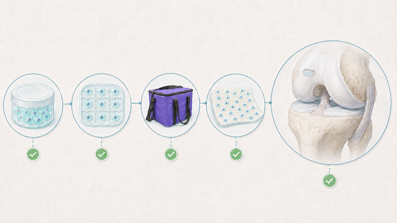
Between the two surgeries, cartilage cells spend approximately four weeks in a place patients do not normally visit – GMP (Good manufacturing practice) laboratory. For patients, this is perhaps the most mysterious stage of the entire treatment. So let us open the laboratory doors and take a closer look at what happens from biopsy collection to cell reimplantation.
What “GMP” Really Means
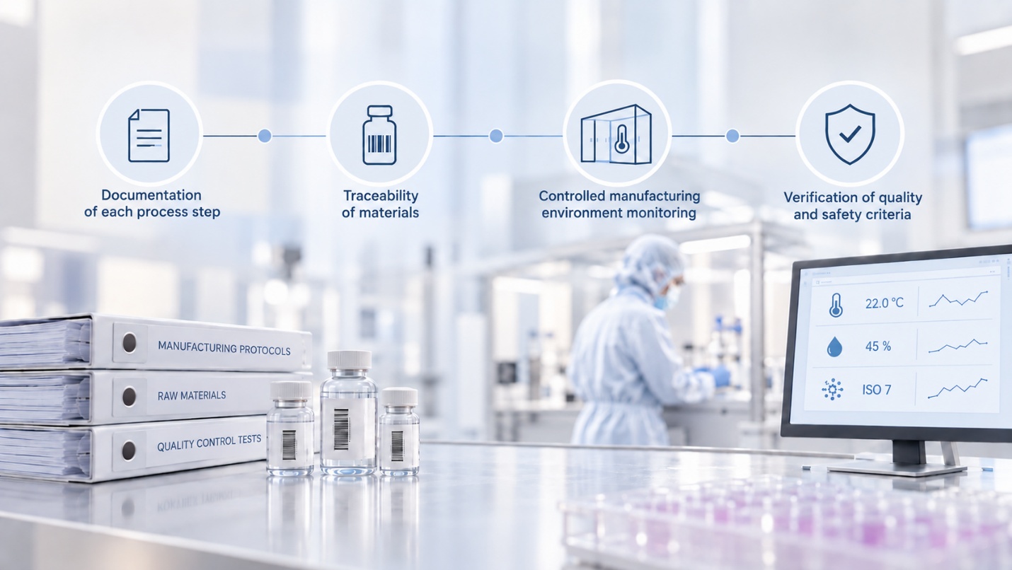
GMP stands for Good Manufacturing Practice, an internationally recognized quality system used in pharmaceutical manufacturing. GMP requirements ensure that every stage of the process is documented, all materials used are traceable, and the manufacturing environment is strictly controlled and continuously monitored.
Prepared cells may be released for use in a patient only after all required batch – release tests have been completed, and the product has been confirmed to meet the established quality and safety criteria. When living cells are used for treatment, this level of discipline ensures that the entire process is carried out consistently, safely, and under controlled conditions.
Step 1: Receiving the Biopsy Sample
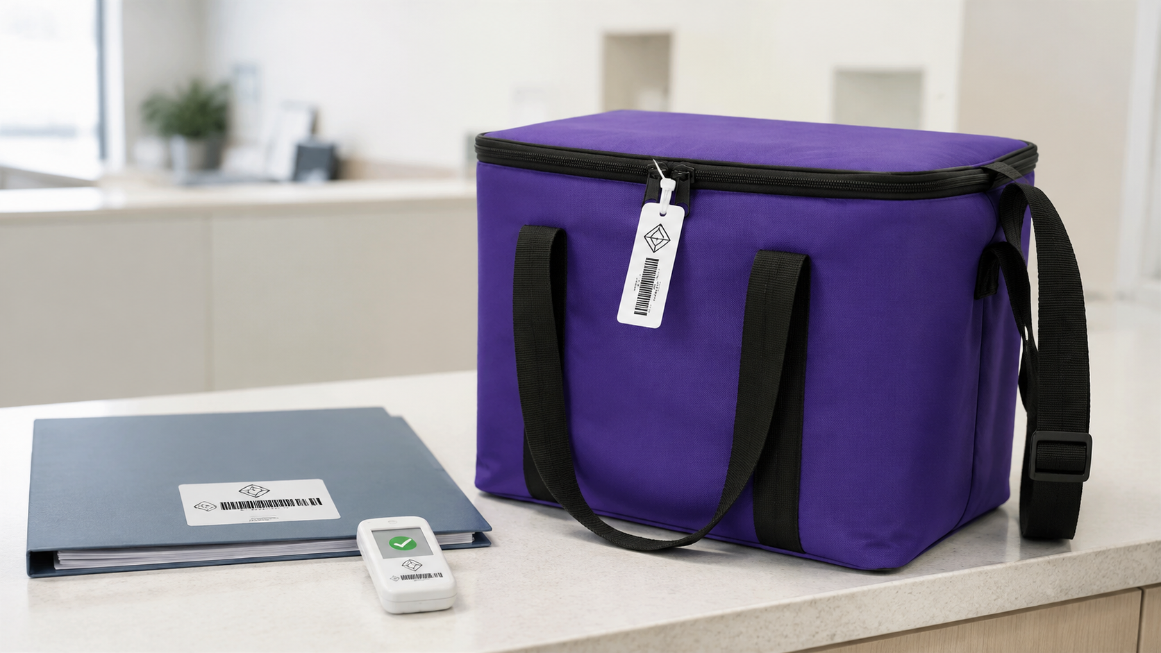
The cartilage sample is transported from the operating room to the laboratory under controlled conditions. Upon arrival, it is registered, its identity is verified, and it is assigned a unique code linked to the batch documentation that accompanies the sample throughout every subsequent stage.
This traceability ensures that, from receipt of the sample through cell implantation, the laboratory is working specifically with your cells. It is one of the most important safety principles in cell therapy.
Step 2: Isolating Your Cells
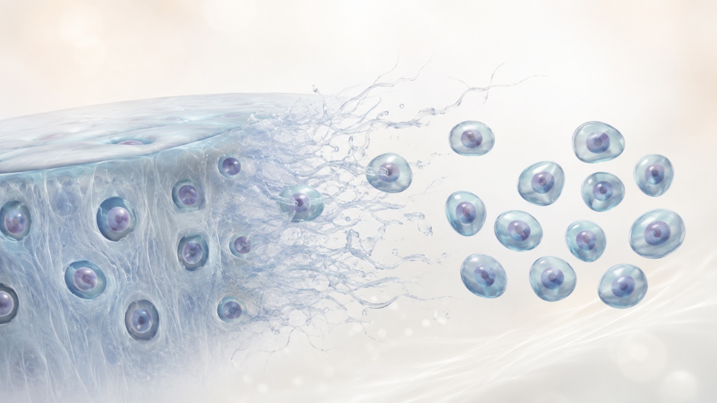
Laboratory specialist’s isolate chondrocytes – the cells that form cartilage tissue – from the cartilage sample. These are the cells that will later be used to repair the damaged cartilage.
The next step is to create suitability, strictly controlled conditions in which the cells can multiply while preserving their viability and the characteristics typical of cartilage tissue.
Step 3: Cell Culture and Expansion (About 4 Weeks)
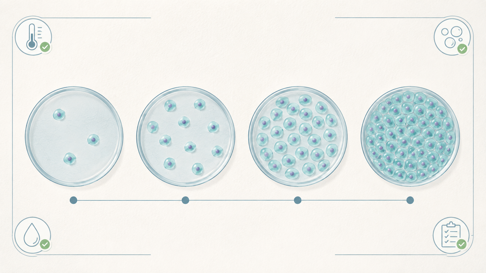
Your cells are cultured under precisely controlled conditions, including temperature, gas composition, and nutrient levels. Over approximately four weeks, the cells isolated from the small biopsy sample are expanded into millions of cells – the quantity required to treat the damaged area of cartilage.
Throughout this period, the process is systematically monitored, supervised, and documented. Cell growth, cell condition, and culture conditions are continuously assessed.
Step 4: Delivery for Implantation
Before your cells are released for implantation, the required quality and safety tests are performed. Only cells that meet all established specifications may be used.
Once the cell preparation has been confirmed as suitable for use, the required cell dose is prepared. The dose is calculated according to the size of the damaged cartilage area.
The preparation is then transported under controlled conditions to the healthcare facility for the second surgery. During the procedure, the cells are seeded onto a collagen membrane, which is subsequently implanted into the damaged area of the knee.
Why This Matters — to Patients and Surgeons Alike
For the patient, this provides reassurance confidence: the weeks spent in the laboratory form part of a systematically controlled, documented, and verified process based on one essential principle – your cells are handled safely and responsibly.
For a surgeon, it answers a fundamental question, even if it is not always asked aloud: can I trust the process to which I am entrusting my patient’s cells? GMP – based manufacturing, complete traceability, and release testing make it possible to use cartilage cell therapy as a controlled innovative clinical procedure.
The science of cartilage cell culture has existed for several decades. However, applying this method reliably to every patient requires strict and consistent manufacturing discipline. This discipline is the foundation of the specialized GMP laboratory’s work.
Frequently Asked Questions
How do you ensure that my cells are not mixed up with another person’s cells?
Complete traceability and identity checks are maintained from the moment the sample is received. All information is carefully recorded in the manufacturing batch documentation.
What is tested before the cells are implanted?
All required quality and safety checks are completed before implantation. The laboratory also confirms that the cells culture is free from contamination and suitable for use.
Why is the date of the second surgery confirmed only later?
The exact date is scheduled only after the laboratory has confirmed that the cells are safe to use. The timing of the surgery therefore depends primarily on the results of the quality and safe checks.
Where is the laboratory located?
The KELI GMP facility in Kaunas, Lithuania.
This article is provided for informational purposes only and is not a substitute for individual medical consultation. Cartilage damage and possible treatment options must be assessed by a qualified orthopaedic surgeon. Each patient’s condition, treatment options, and recovery may differ.
→ Find out if you are a candidate for RECARTA — take the eligibility review at recarta.eu
About the Author
Justinas Maciulaitis, MD, PhD
Justinas Mačiulaitis, MD, PhD, is an orthopaedic surgeon and translational research specialist with more than 15 years of experience in regenerative medicine. He is the founder and CEO of KELI Therapeutics, an international advanced biotechnology company developing and manufacturing cell therapies for cartilage regeneration. His doctoral dissertation in translational regenerative medicine, completed at the Lithuanian University of Health Sciences, contributed to the development of RECARTA—a GMP-manufactured autologous and allogeneic chondrocyte therapy intended for the treatment of focal knee cartilage defects. Dr Mačiulaitis has presented at international scientific conferences and contributed to translational research in cartilage repair, inflammation management, and cell therapy manufacturing.
Sources
- European Medicines Agency. Good Manufacturing Practice. ema.europa.eu
- Brittberg M et al. Treatment of deep cartilage defects in the knee with autologous chondrocyte transplantation. N Engl J Med. 1994;331(14):889–895.
- Ebert JR et al. 10-Year Prospective Clinical and Radiological Evaluation After Matrix-Induced Autologous Chondrocyte Implantation. Am J Sports Med. 2024.
Tags
More Articles
Hyaline vs Fibrocartilage: Why Does Tissue Quality Matter So Much?
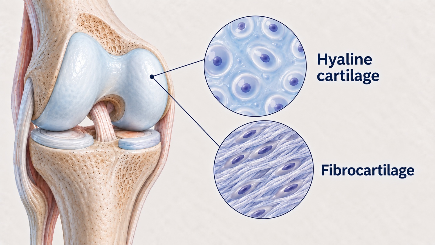
Two patients may undergo seemingly identical cartilage repair procedures, yet five years later their outcomes may be entirely different. This difference is often determined not by the surgeon who performed the procedure or by the patient’s efforts during rehabilitation, but by the type of tissue that formed after surgery. The damaged area may be repaired with hyaline cartilage of fibrocartilage. Although both are considered cartilage tissue, their structure, mechanical properties, and long – term durability differ significantly. This difference often determines whether the joint continues to function smoothly for many years or whether symptoms eventually return.
Stage 1, Stage 2: The Complete RECARTA Journey
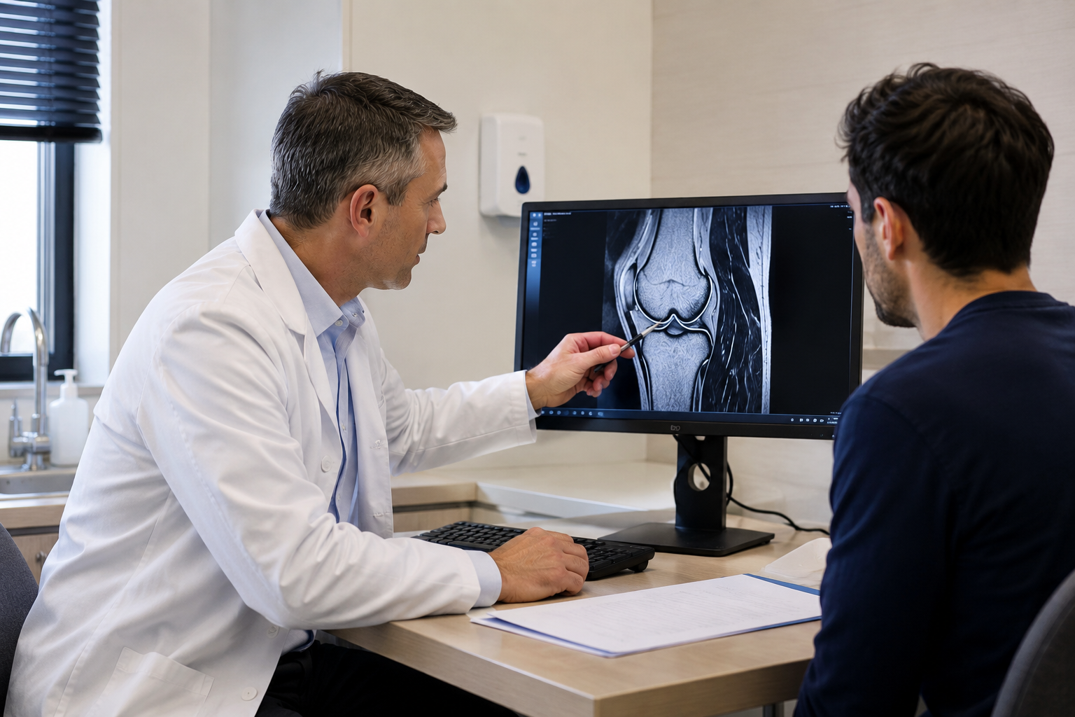
When considering cartilage cell therapy, the greatest concern is often not the treatment itself, but the uncertainty. Two surgeries instead of one, waiting in between, and the process taking place in a specialized laboratory that the patient does not see. When you don't know what's happening at each stage, doubting and worrying is completely natural. Therefore, we present the entire RECARTA treatment process – clearly and consistently, from the first consultation to rehabilitation.