
Stage 1, Stage 2: The Complete RECARTA Journey
When considering cartilage cell therapy, the greatest concern is often not the treatment itself, but the uncertainty. Two surgeries instead of one, waiting in between, and the process taking place in a specialized laboratory that the patient does not see. When you don't know what's happening at each stage, doubting and worrying is completely natural. Therefore, we present the entire RECARTA treatment process – clearly and consistently, from the first consultation to rehabilitation.
Before Anything: The Consultation
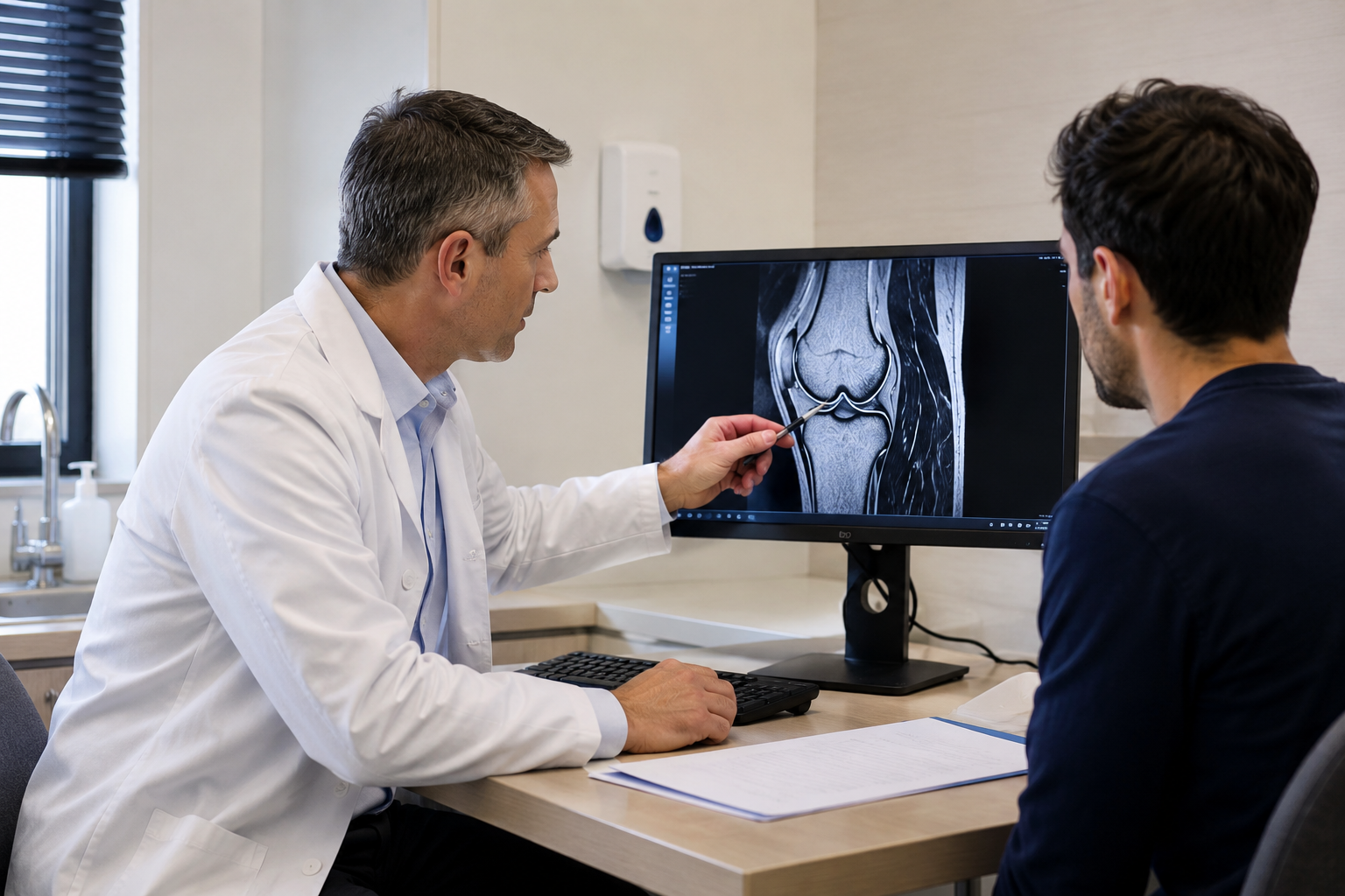
It all starts with a consultation with an orthopedic surgeon. During it, your symptoms, X-ray and magnetic resonance imaging (MRI) results, and the overall condition of your knee joint are assessed. Cartilage cell therapy is not suitable for every patient. It is intended for focal cartilage defects – specific, clearly defined lesion sites, usually classified as ICRS grade III or IV, where other knee structures are stable. If the surgeon confirms that this treatment method is suitable for you, a two-stage process begins.
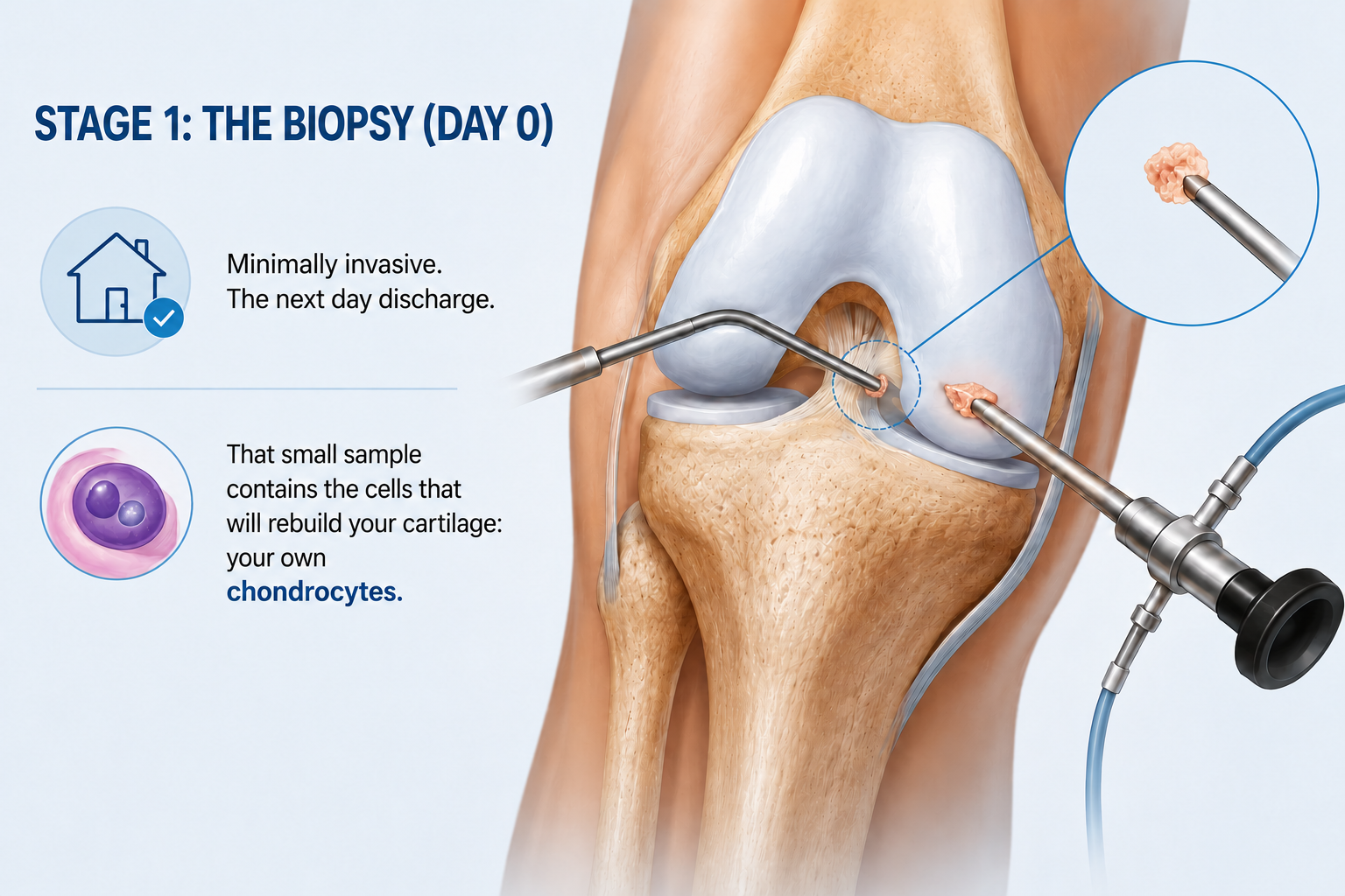
Stage 1: The Biopsy (Day 0)
The first procedure is short and minimally invasive. When performing arthroscopy – surgery through small incisions – the surgeon takes a small sample of healthy cartilage from the unloaded area of the knee joint. It is about the size of a pen tip. Usually, the patient can go home the very next day.
This small sample contains your chondrocytes – cartilage cells that will later be cultured in the facility and used to restore the damaged area.
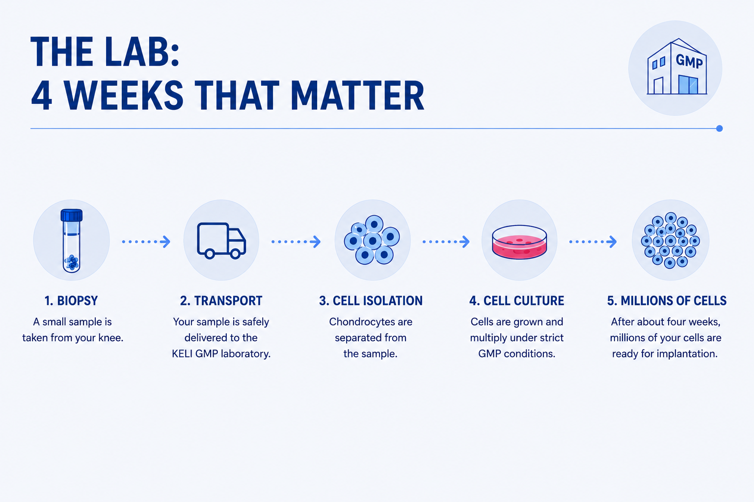
The specialized lab: 4 Weeks That Matter
The biopsy sample is delivered to KELI Therapeutics’ licensed GMP facility in Kaunas, Lithuania. GMP is Good Manufacturing Practice, a high-quality standard applied in pharmaceutical and biotechnological manufacturing. In the facility, chondrocytes are isolated from the collected cartilage sample and grown under strictly controlled conditions. In about four weeks, millions of cells needed to treat the cartilage defect are grown.
This part of the treatment often raises the most questions for patients because the whole process takes place in the laboratory. RECARTA can be delivered using one of two types of cells: your own (autologous) or those of a carefully screened donor (allogeneic). Both use living cartilage cells and are prepared in a certified laboratory under the same rigorous quality standards - the only difference is where the cells originate. Which option is more suitable depends on your clinical situation, and this is decided together with your doctor on an individual basis. Whichever route is chosen, the laboratory provides the cells with the conditions they need to multiply until there are enough to repair the cartilage defect.
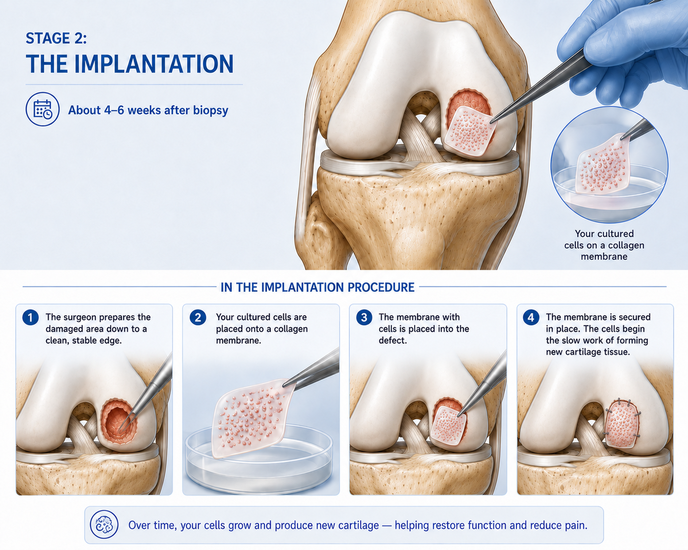
Stage 2: The Implantation (4–6 Weeks After Biopsy)
Once the cells are grown and quality control tests confirm they can be used, a second operation is planned. The exact date is confirmed by the production team upon receiving the final test results. Implantation is usually performed four weeks after the biopsy.
During the operation, the surgeon prepares the damaged cartilage site by creating clean and stable defect edges. Then the cells grown in the certified manufacturing facility are placed on the special collagen membrane and secured in the prepared area of the cartilage defect. From this moment, a process of new cartilage tissue formation and maturation begins.
After: Rehabilitation Is Not Optional — It Is Half the Result
Implantation is not the end of treatment. Initially, the forming tissue is fragile, so it needs time to mature and strengthen. During this period, consistent, specialist - supervised rehabilitation is especially important:
- Weeks 0–6: Protected weight-bearing, controlled movement, reducing swelling.
- Weeks 6–12: Gradual return to everyday activity.
- Months 3–6: Strength building; most patients return to normal daily life.
- Months 6–12: Progressive loading and, for many, a return to sport.
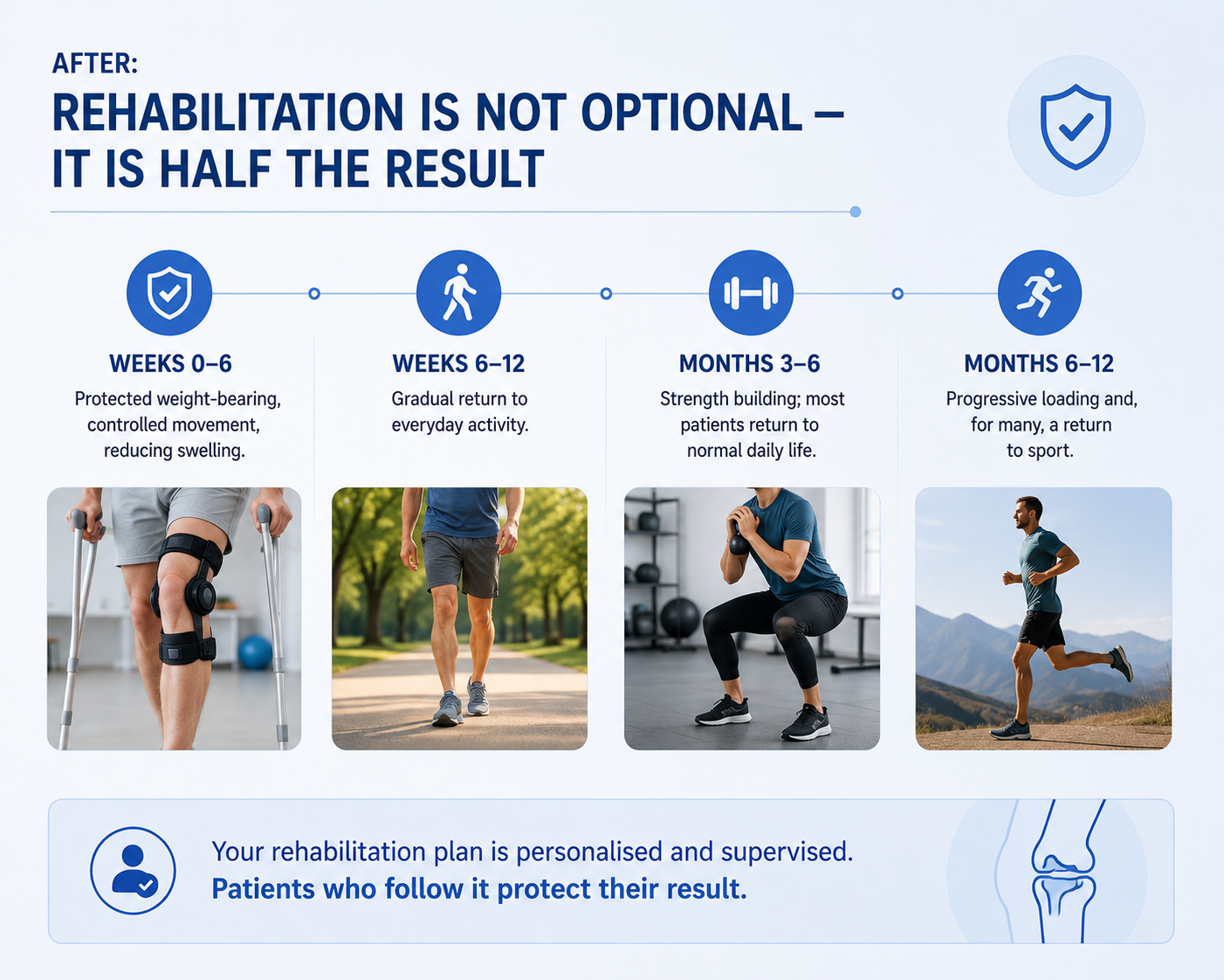
An individual rehabilitation plan is created for each patient. Its course depends on the size and location of the cartilage defect, the surgery performed, and the overall condition of the knee joint. Consistent adherence to the plan is one of the most important conditions for successful treatment.
Why the Two Stages Are Worth It
One procedure would be simpler, but it is precisely the two-step method that allows the use of live cells from your own cartilage for treatment. The goal of this treatment is to form cartilage tissue that is more similar in properties to natural cartilage rather than scar tissue, which can develop with some other procedures. The clinical data supporting this method have been collected for more than 30 years, and the observation period for published long-term studies is 10 years.
Two procedures. There are four weeks between them. During that time, your own cells are grown in a certified manufacturing facility and then used to restore the damaged knee cartilage.
Frequently Asked Questions
How long between the two surgeries? Usually four weeks. The precise date is set together with a treating orthopedic surgeon.
Is the biopsy painful? It is a short keyhole procedure. Most patients go home the next day with mild discomfort.
Whose cells are used? Only your own. The manufacturing facility multiplies the cells taken from your biopsy.
When can I walk normally again? Most patients return to everyday activity within about 12 weeks, following their rehabilitation plan.
This article is for informational purposes only and does not constitute medical advice. All cartilage conditions should be assessed by a qualified orthopedic specialist. Individual presentations and treatment options vary.
→ Ready to find out if RECARTA is right for your knee? Take the eligibility check at recarta.eu.
About the Author
Justinas Maciulaitis, MD, PhD
Justinas Maciulaitis, MD, PhD — an orthopedic surgeon and translational research specialist with more than 15 years of experience in regenerative medicine. He is the founder and chief executive of KELI Therapeutics — an international advanced biotechnology company specializing in the development and manufacture of cell therapies for cartilage regeneration. His doctoral dissertation in translational regenerative medicine at the Lithuanian University of Health Sciences contributed to the development of RECARTA from donor cells (allogeneic RECARTA). RECARTA from donor cells is planned to be available from early 2027. Dr. Maciulaitis has presented at international scientific conferences and contributed to translational research in the areas of cartilage repair, inflammation management, and cell therapy manufacturing.
Sources
-
International Cartilage Repair Society. ICRS Cartilage Injury Evaluation Package. cartilage.org
-
Brittberg M et al. Treatment of deep cartilage defects in the knee with autologous chondrocyte transplantation. N Engl J Med. 1994;331(14):889–895.
-
Ebert JR et al. 10-Year Prospective Clinical and Radiological Evaluation After Matrix-Induced Autologous Chondrocyte Implantation. Am J Sports Med. 2024.
Tags
More Articles
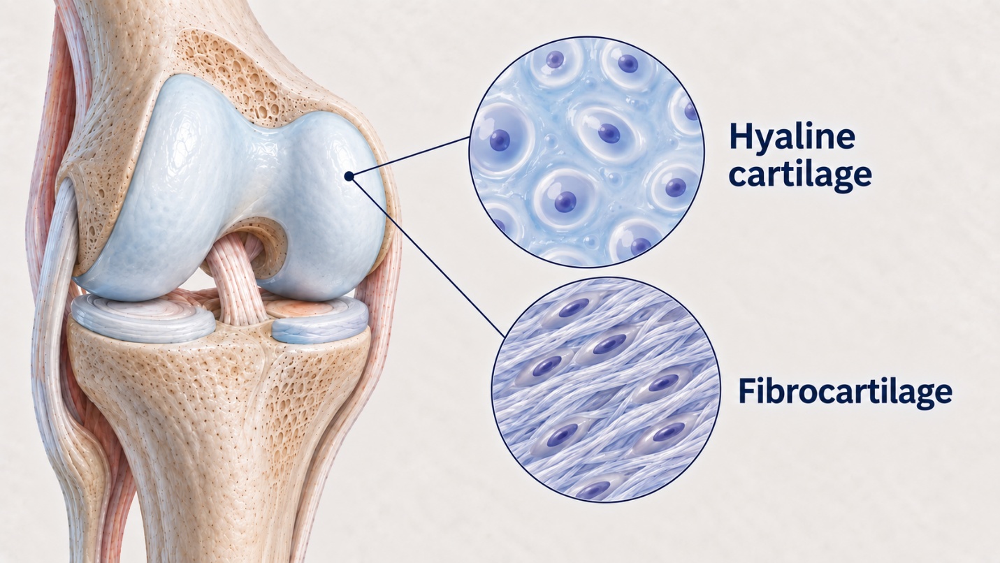
Hyaline vs Fibrocartilage: Why Does Tissue Quality Matter So Much?
Two patients may undergo seemingly identical cartilage repair procedures, yet five years later their outcomes may be entirely different. This difference is often determined not by the surgeon who performed the procedure or by the patient’s efforts during rehabilitation, but by the type of tissue that formed after surgery. The damaged area may be repaired with hyaline cartilage of fibrocartilage. Although both are considered cartilage tissue, their structure, mechanical properties, and long – term durability differ significantly. This difference often determines whether the joint continues to function smoothly for many years or whether symptoms eventually return.
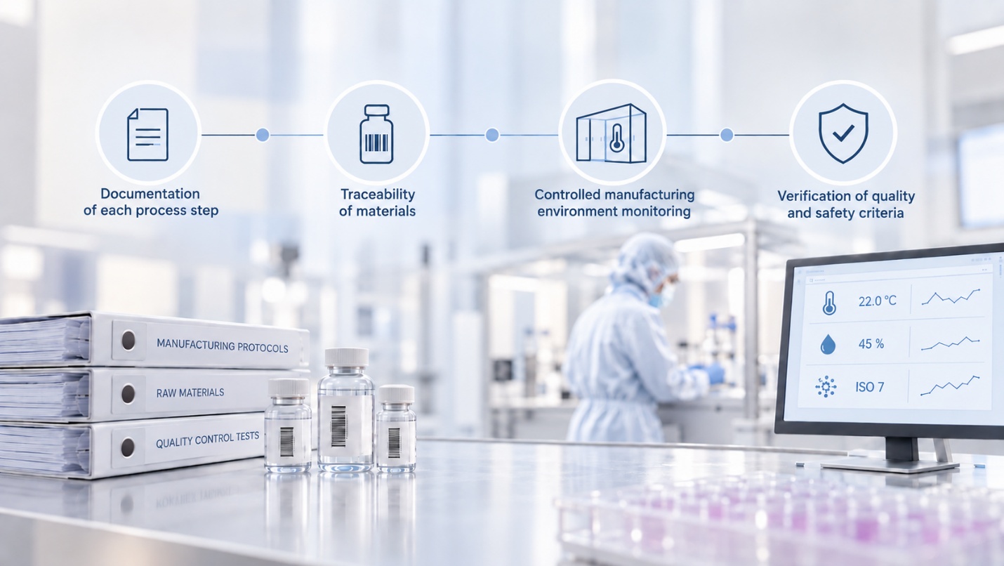
Inside Our GMP Lab: From Biopsy to Reimplantation
Between the two surgeries, cartilage cells spend approximately four weeks in a place patients do not normally visit – GMP (Good manufacturing practice) laboratory. For patients, this is perhaps the most mysterious stage of the entire treatment. So let us open the laboratory doors and take a closer look at what happens from biopsy collection to cell reimplantation.