
Microfracture Failed. What Now?
You had the microfracture procedure. For a year, or perhaps even two, your knee felt better, but over time the pain gradually began to return. During a consultation, you may have been told, “There isn’t much more we can do.” However, this is not necessarily true.
When the benefits of a microfracture procedure begin to wear off, treatment options do not simply disappear. For many patients, this may not be the end of their treatment journey, but rather the beginning of a more focused discussion about the next steps.

Why the Effects of Microfracture May Weaken Over Time?
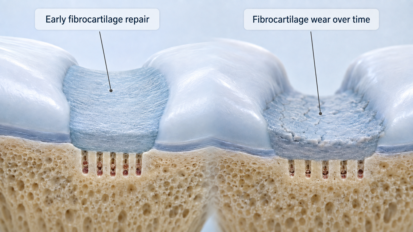
In a microfracture procedure, small openings are made in the bone beneath the damaged cartilage, reaching into the bone-marrow cavity. Through these, blood-forming cells reach the cartilage defect and help form a reparative, cartilage-like tissue. It is a relatively quick and inexpensive procedure, suited to treating small cartilage lesions. Over time, however, its effect weakens for some patients — and there are several reasons why.
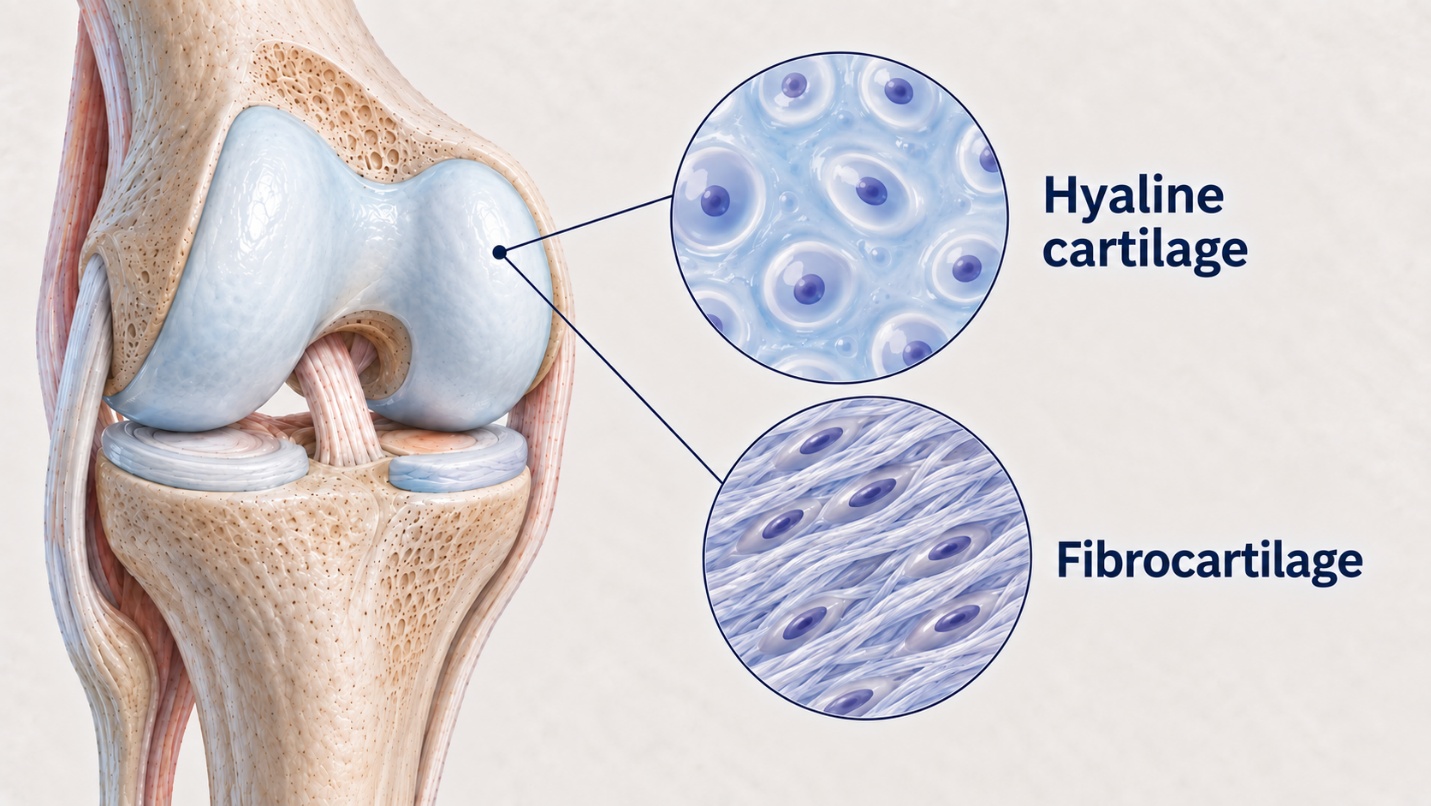
1. Quality of the repair tissue. The tissue formed after microfracture is mainly fibrocartilage — a scar-like structure. It is not the same as the smooth, durable hyaline cartilage that naturally lines the surfaces of a healthy knee joint. Fibrocartilage is mechanically weaker and more prone to wear.
2. Progressive deterioration. Clinical studies show that, while microfracture can provide short-term relief, the repair tissue often begins to break down within a few years (more than 60 % of patients). For some patients, deterioration becomes noticeable at around the two-year mark — at which point the earlier pain can return, along with the need for further treatment.
3. The mechanical demands on the joint. Everyday activity places heavy demands on the knee. Walking alone involves roughly 2–3 million steps a year, and the load on the joint can reach up to five times body weight. Pivoting, jumping, and high-impact sport amplify these forces further. When the tissue formed after microfracture cannot withstand such loads, the bone beneath the cartilage can become exposed — intensifying both pain and loss of joint function.
4. The limits of natural healing. Microfracture relies on the body's natural capacity to heal, but for repairing cartilage this capacity is often not enough on its own. Without an additional solution, the body struggles to meet the demands placed on a weight-bearing joint.
This is not your fault. It is a known limitation of this treatment method, particularly when the cartilage defect is larger than about 2 cm². In such cases, the long-term results of microfracture are not always reliable.
Why It Helps Not to Delay the Decision
There is one more reason to consider your options early. Microfracture damages the subchondral bone — the foundation on which new cartilage forms. Long-term research shows that when microfracture does not provide a lasting result, later cartilage restoration procedures such as cell therapy tend to be less successful at those previously treated sites than in untouched cartilage. In other words, an unsuccessful microfracture can make subsequent treatment harder.
This does not mean cell therapy is no longer possible after microfracture — many such patients are still treated successfully. But it does mean that the earlier the right treatment is chosen, the better the long-term outlook.
What Does an Unsuccessful Microfracture Procedure Actually Mean?
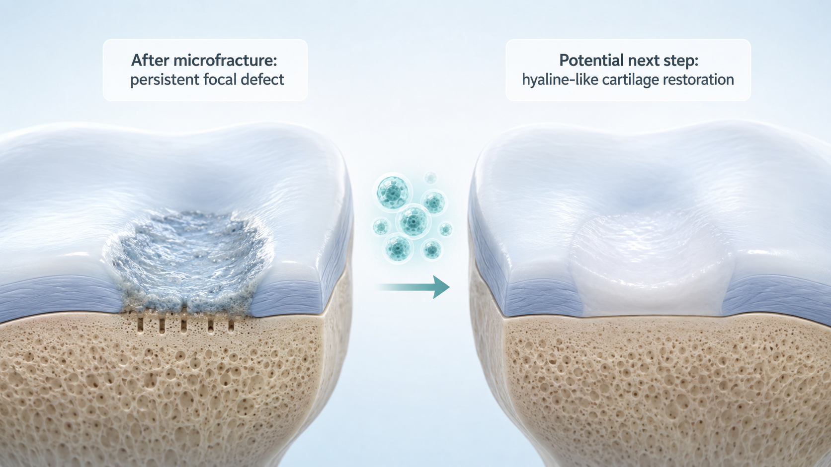
In most cases, it means that a focal cartilage defect remains in the knee joint. This may indicate that treatment should be considered with the aim of forming tissue that is not scar-like but more closely resembles hyaline cartilage in its biological properties. Importantly, a previous microfracture procedure does not rule out cell therapy. Potential candidates for RECARTA include active adults whose initial procedure did not provide lasting results.
When May Cell Therapy Be Used?
RECARTA uses the patient’s own cartilage cells. These cells are expanded in a laboratory that complies with Good Manufacturing Practice (GMP) requirements and are later implanted into the cartilage defect. The aim of this treatment is to form tissue that more closely resembles natural cartilage in its biological properties and can withstand the loads placed on the joint for many years rather than just a few months. The principle of autologous cartilage cell therapy has been used in clinical practice for more than 30 years, and published patient follow-up data cover periods of up to 10 years.
For a patient whose microfracture benefits have diminished over time, the treatment pathway may be understood as follows: the tissue formed after the initial procedure was not sufficiently durable, so the next step may be a treatment designed to produce stronger tissue that more closely resembles natural cartilage.
What Should You Do Next?
If the benefits of the microfracture procedure have diminished, consider taking these three steps:
- Repeat the MRI scan — An MRI performed before the microfracture procedure is not sufficient to assess the current condition of your knee.
- Consult an orthopedic surgeon: Find out the size of the cartilage defect and whether the knee joint is stable.
- Discuss your treatment options: if the defect is focal, larger than approximately 2 cm², and the knee joint is stable, you may be a suitable candidate for further treatment.
Sometimes the statement “nothing more can be done” may indeed be justified. More often, however, it means that nothing more can be done using the same treatment method.
Frequently Asked Questions
Can cell therapy be used after a microfracture procedure?
Often, yes. A previous microfracture procedure does not rule out cell therapy. However, eligibility for treatment is assessed individually in each case.
Why did the benefits of the microfracture procedure wear off?
Microfracture usually produces fibrocartilage, which resembles scar tissue. It is mechanically weaker than natural hyaline cartilage and may therefore be worn down over time, particularly when treating a larger cartilage defect.
How long should you wait after an unsuccessful microfracture procedure?
There is no single rule that applies to every patient. An MRI scan and a consultation with an orthopedic surgeon will help determine the most appropriate time for further treatment.
Is a second procedure riskier?
This depends on the individual condition of the knee joint, the remaining cartilage defect, previous treatment, and other factors. Your doctor will assess these circumstances and discuss the potential benefits and risks with you.
This article is for informational purposes only and does not constitute medical advice. All cartilage conditions should be assessed by a qualified orthopaedic specialist. Individual presentations and treatment options vary.
→ Had a microfracture that did not hold? Find out if RECARTA is the right next step — take the eligibility review at recarta.eu
About the Author
Justinas Maciulaitis, MD, PhD
Justinas Maciulaitis, MD, PhD — an orthopedic surgeon and translational research specialist with more than 15 years of experience in regenerative medicine. He is the founder and chief executive of KELI Therapeutics — an international advanced biotechnology company specializing in the development and manufacture of cell therapies for cartilage regeneration. His doctoral dissertation in translational regenerative medicine at the Lithuanian University of Health Sciences led to the development of RECARTA — an autologous and allogeneic chondrocyte therapy manufactured under Good Manufacturing Practice conditions, designed to treat focal cartilage defects of the knee. Dr. Maciulaitis has presented at international scientific conferences and contributed to translational research in the areas of cartilage repair, inflammation management, and cell therapy manufacturing.
Sources
- Brittberg M et al. Treatment of deep cartilage defects in the knee with autologous chondrocyte transplantation. N Engl J Med. 1994;331(14):889–895.
- Kon E et al. ACI versus microfracture: a 10-year follow-up. Knee Surg Sports Traumatol Arthrosc. 2012.
- Ebert JR et al. 10-Year Prospective Clinical and Radiological Evaluation After Matrix-Induced Autologous Chondrocyte Implantation. Am J Sports Med. 2024.
- International Cartilage Repair Society. ICRS Cartilage Injury Evaluation Package. cartilage.org
- Merkely G, Ogura T, Bryant T, Minas T. Severe Bone Marrow Edema Among Patients Who Underwent Prior Marrow Stimulation Technique Is a Significant Predictor of Graft Failure After Autologous Chondrocyte Implantation. Am J Sports Med. 2019;47(8):1874–1884.
- Cogan CJ, Friedman J, You J, et al. Prior Bone Marrow Stimulation Surgery Influences Outcomes After Cell-Based Cartilage Restoration: A Systematic Review and Meta-analysis. Orthop J Sports Med. 2021;9(9):23259671211035384.
Tags
More Articles
Hyaline vs Fibrocartilage: Why Does Tissue Quality Matter So Much?
Two patients may undergo seemingly identical cartilage repair procedures, yet five years later their outcomes may be entirely different. This difference is often determined not by the surgeon who performed the procedure or by the patient’s efforts during rehabilitation, but by the type of tissue that formed after surgery. The damaged area may be repaired with hyaline cartilage of fibrocartilage. Although both are considered cartilage tissue, their structure, mechanical properties, and long – term durability differ significantly. This difference often determines whether the joint continues to function smoothly for many years or whether symptoms eventually return.
Inside Our GMP Lab: From Biopsy to Reimplantation
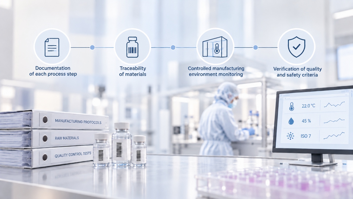
Between the two surgeries, cartilage cells spend approximately four weeks in a place patients do not normally visit – GMP (Good manufacturing practice) laboratory. For patients, this is perhaps the most mysterious stage of the entire treatment. So let us open the laboratory doors and take a closer look at what happens from biopsy collection to cell reimplantation.